Severity: 8192
Message: str_replace(): Passing null to parameter #3 ($subject) of type array|string is deprecated
Filename: front/sub-sub-category.php
Line Number: 200
Backtrace:
File: /home2/psawcmdw/public_html/www.psawlaboratoryequipments.com/application/views/template/front/sub-sub-category.php
Line: 200
Function: str_replace
File: /home2/psawcmdw/public_html/www.psawlaboratoryequipments.com/application/views/template.php
Line: 13
Function: view
File: /home2/psawcmdw/public_html/www.psawlaboratoryequipments.com/application/core/MY_Controller.php
Line: 26
Function: view
File: /home2/psawcmdw/public_html/www.psawlaboratoryequipments.com/application/controllers/Welcome.php
Line: 105
Function: get_user_template
File: /home2/psawcmdw/public_html/www.psawlaboratoryequipments.com/index.php
Line: 315
Function: require_once
Severity: 8192
Message: str_replace(): Passing null to parameter #3 ($subject) of type array|string is deprecated
Filename: front/sub-sub-category.php
Line Number: 201
Backtrace:
File: /home2/psawcmdw/public_html/www.psawlaboratoryequipments.com/application/views/template/front/sub-sub-category.php
Line: 201
Function: str_replace
File: /home2/psawcmdw/public_html/www.psawlaboratoryequipments.com/application/views/template.php
Line: 13
Function: view
File: /home2/psawcmdw/public_html/www.psawlaboratoryequipments.com/application/core/MY_Controller.php
Line: 26
Function: view
File: /home2/psawcmdw/public_html/www.psawlaboratoryequipments.com/application/controllers/Welcome.php
Line: 105
Function: get_user_template
File: /home2/psawcmdw/public_html/www.psawlaboratoryequipments.com/index.php
Line: 315
Function: require_once
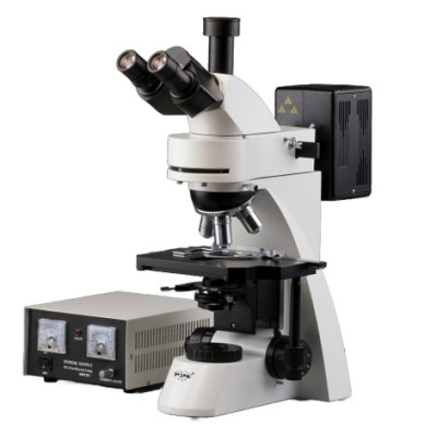
The Global Standard for Molecular Imaging. PSAW is the leading Fluorescent Research Microscope Manufacturer in India, delivering high-sensitivity optical systems for genetics, immunology, and oncology.
As the premier Fluorescent Research Microscope Manufacturer in India, PSAW redefines the limits of cellular observation. Our Fluorescent series (PSAW-102 & 106) utilizes advanced Mercury Vapor or High-Intensity LED light sources to excite fluorophores with extreme precision. These systems are engineered to capture low-energy light emissions, allowing researchers to track targeted DNA sequences and proteins against an ultra-dark background.
We are recognized as a trusted Fluorescent Research Microscope Supplier in India, providing modular trinocular configurations to medical colleges, biotech firms, and government research centers. Our role as a global Exporter ensures that labs from Europe to Southeast Asia receive high-numerical aperture (NA) objectives and specialized dichroic mirror systems optimized for FISH, multi-color imaging, and neurodegenerative studies.
As a top Supplier in India, we provide systems for Fluorescence In Situ Hybridization (FISH) to detect chromosomal abnormalities in cancer research.
Widely utilized by our Export partners for detecting pathogens like Mycobacterium tuberculosis with extreme specificity and speed.
Used in water quality analysis to identify autofluorescent algae and organic pollutants, supplied by PSAW as a leading Manufacturer.
The operational power of PSAW Fluorescent systems lies in their extraordinary signal-to-noise ratio. By isolating specific cellular components without staining the surrounding tissue, researchers gain a deeper understanding of subcellular interactions. As an Exporter of high-precision optics, we integrate high-numerical aperture (NA) objectives that maximize light collection—essential for capturing faint fluorescent signals in dynamic biological processes. This makes our microscopes indispensable for both routine clinical screening and multi-color academic research.
We combine indigenous manufacturing excellence with international grade filter cubes and high-intensity excitation systems, offering 4K imaging compatibility for modern digital laboratories.
Yes, as a specialized Supplier in India, we provide sets for DAPI, FITC, TRITC, and Texas Red, allowing for multiplexing in oncology and developmental biology.
Every unit exported by PSAW undergoes strict UV-leakage testing and is shipped in reinforced, moisture-sealed packaging to preserve the integrity of the dichroic mirrors.
PSAW is a premier Fluorescent Research Microscope Exporter to: USA, UK, Germany, Singapore, UAE, South Africa, Russia, Brazil, Malaysia, Vietnam, and 70+ countries.
Top Fluorescent Research Microscope Supplier in India serving hubs like Mumbai, Bangalore, Delhi NCR, Hyderabad, Chennai, Pune, Kolkata, and Chandigarh.
© PSAW Laboratory Equipments - Official Manufacturer, Supplier & Exporter of Advanced Fluorescence Systems.